Embolism Diagnosis: How Doctors Find Dangerous Clots
Sudden shortness of breath or sharp chest pain can mean a blood clot has moved into your lungs. That’s a pulmonary embolism (PE), and it can be life‑threatening. Knowing what doctors look for helps you get the right care fast.
First step: the clinical check. The medical team will ask about recent surgery, long travel, cancer, hormones, or leg swelling. They’ll measure oxygen levels, heart rate, blood pressure, and listen to your chest. These quick checks tell them how urgent the situation is.
Key tests you’ll meet in the ER
D-dimer is a blood test often done first. A low D-dimer makes a clot unlikely, so no scary scans for most low-risk people. But a high D-dimer doesn’t prove a clot — it just prompts more testing.
Compression ultrasound of the legs looks for deep vein thrombosis (DVT). If a DVT is found, doctors may treat you for embolism even if lung imaging isn’t done right away. The ultrasound is quick and safe, and it’s the main test when symptoms point to a leg clot.
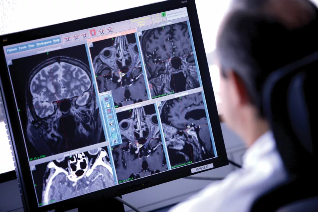
CT pulmonary angiography (CTPA) is the most common way to confirm a pulmonary embolism. It uses contrast dye and shows clots inside lung arteries. It’s fast and accurate but not ideal if you have poor kidney function or contrast allergies.
Ventilation‑perfusion (V/Q) scan is an alternative when CT isn’t safe — for example during pregnancy or severe kidney problems. V/Q scans compare air flow and blood flow in the lungs to spot mismatches that suggest a clot.
Other useful checks
Electrocardiogram (ECG) and chest X‑ray help rule out other causes of chest pain. Echocardiography checks how the right heart is coping — big clots can strain the heart and change treatment choices.
Sometimes doctors use formal scoring systems like the Wells score or Geneva score. These tools combine symptoms and signs to estimate the chance of PE and guide whether testing is needed now or later.
What happens after diagnosis matters. If tests confirm a clot, anticoagulant (blood thinner) treatment usually starts quickly. The type, dose, and length of treatment depend on cause, location, and bleeding risks.
When should you act now? Call emergency services for sudden severe shortness of breath, fainting, heavy chest pain, or collapsing. Don’t wait for scheduled visits if you feel acutely worse.
Practical tip: bring a list of current medications, recent surgeries, and any history of clots when you go to the ER. That helps speed up correct testing and treatment.
Knowing the tests and steps helps you feel less lost if a clot is suspected. Talk openly with your care team about risks, test options, and next steps — it makes the process clearer and faster.
The Role of CT Scans in Embolism Diagnosis and Management
As a blogger, I've recently been researching the role of CT scans in embolism diagnosis and management. It's fascinating to learn how crucial these scans have become in accurately detecting blood clots, especially those in the lungs and brain. The speed and precision of CT scans allow doctors to make swift decisions on the appropriate treatment for patients, which can be lifesaving in many cases. Furthermore, CT scans can also help monitor the effectiveness of ongoing treatments for embolisms. Overall, it's clear that CT scans play a vital part in the early detection and management of embolisms, ultimately improving patient outcomes.
read more