Understanding Embolism and the Need for Accurate Diagnosis
An embolism is a serious medical condition characterized by the sudden blockage of a blood vessel, usually by a blood clot or an air bubble. This blockage can lead to severe complications, such as stroke, heart attack, or even death, if not diagnosed and treated promptly. As a blogger who is passionate about raising awareness about health issues, I aim to shed light on the critical role of CT scans in the diagnosis and management of embolism. In this article, I will discuss different aspects of embolism diagnosis and management using CT scans, with a focus on their accuracy and reliability.
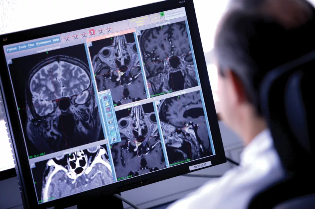
CT Scans: A Powerful Tool for Detecting Embolism
Computed tomography (CT) scans are a vital diagnostic tool in the detection of embolism. This non-invasive imaging technique uses X-rays to create detailed cross-sectional images of the body's internal structures, allowing doctors to visualize the presence and location of blood clots or air bubbles causing the blockage. CT scans have revolutionized the diagnosis of embolism, as they provide a quick, accurate, and safe means of identifying the source of the problem and planning the most effective course of treatment.
Types of CT Scans for Embolism Diagnosis
There are several types of CT scans used for diagnosing embolism, depending on the location of the suspected blockage. For instance, a pulmonary CT angiography (CTA) is used to detect blood clots in the lungs, while a cerebral CTA helps identify blockages in the blood vessels of the brain. Additionally, a CT venography (CTV) can be used to visualize veins, particularly in the lower extremities, where deep vein thrombosis (DVT) may occur. The choice of CT scan type depends on the patient's symptoms and the physician's clinical suspicion.
CT Scans Versus Other Diagnostic Methods
While there are several diagnostic methods for detecting embolism, such as ultrasound, magnetic resonance imaging (MRI), and venography, CT scans offer several advantages. They are faster than most other imaging techniques, allowing for quicker diagnosis and initiation of treatment. Furthermore, CT scans provide high-resolution images that enable physicians to accurately pinpoint the location and size of the blockage. This precision is crucial in determining the most appropriate treatment strategy for each patient.
Risks and Limitations of CT Scans in Embolism Diagnosis
Although CT scans play a crucial role in embolism diagnosis, they are not without risks and limitations. The use of ionizing radiation in CT scans raises concerns about potential harm to the patient, particularly in cases where multiple scans are required. Moreover, CT scans may not always be suitable for certain patients, such as pregnant women or individuals with allergies to contrast agents used in CTA and CTV. In these cases, alternative imaging techniques, like MRI or ultrasound, may be considered.
Embolism Management: How CT Scans Guide Treatment
Once an embolism has been diagnosed using a CT scan, the information obtained from the images plays a critical role in guiding treatment. The location and size of the blockage help physicians determine the most suitable course of action, which may include medication, minimally invasive procedures, or surgery. In some cases, CT scans can also be used to monitor the effectiveness of treatment, such as the dissolution of a blood clot following the administration of clot-busting drugs.
Preventing Embolism: The Value of Early Detection
Preventing embolism is of utmost importance, as the complications associated with this condition can be life-threatening. CT scans serve as a valuable tool for early detection, allowing physicians to identify and treat potential blockages before they cause severe harm. By raising awareness about the importance of early diagnosis and regular check-ups, we can help reduce the incidence of embolism and improve the overall health of our communities.
Conclusion: The Critical Role of CT Scans in Embolism Diagnosis and Management
In conclusion, CT scans play an indispensable role in the accurate and timely diagnosis of embolism, as well as in guiding effective treatment strategies. While there are risks and limitations associated with this imaging technique, the benefits of early detection and accurate diagnosis far outweigh the potential drawbacks. As a blogger committed to educating the public about health issues, I am dedicated to spreading awareness about the importance of CT scans in embolism diagnosis and management, and advocating for their continued use in medical practice.

Darren Ramowski
My name is Calem Goodridge, and I am a pharmaceutical expert with a passion for educating others about medication and diseases. I have dedicated my career to researching and developing life-changing medical treatments. In my spare time, I enjoy writing articles and sharing my knowledge with the general public to help them make informed healthcare decisions. I am also an advocate for safe medication practices and believe that patient education is key to preventing adverse drug events. With my extensive experience in the pharmaceutical industry, I strive to make a positive impact on the lives of others through my writing.
Oh great, another love letter to CT scans-because we definitely needed more radiation in our lives. If you think the speed and detail are a miracle, maybe you’ve never tried an MRI that doesn’t scream “I’m here to save the day”.
Sure, CTs are the superhero of imaging, swooping in with X‑ray powers. Meanwhile, the poor ultrasound sits in the corner like an under‑appreciated sidekick.
CT scans are just a sneaky way for the pharma industry to push more equipment. They claim you need them for every little clot. In reality the radiation dose adds up over time. People think the images are flawless. But every scan has a margin of error. The contrast agents can cause allergic reactions. Some hospitals hide the cost from patients. The push for CTs ignores cheaper ultrasound options. The data from older studies is being cherry‑picked. There’s a lobby that funds the research. If you look at the raw numbers you see a bias. The technology is not as bullet‑proof as they say. Some radiologists even admit they miss small emboli. The rush to diagnose can lead to overtreatment. So next time you hear about a CT ask who profits 😒
Honestly the article is like a textbook rewrite but with way too many buzzwords. It sounds fancy but the core ideas are just the same old stuff we’ve heard a thousand times, and yeah the spelling errors like definately and recieve just prove it.
We don’t need foreign tech dictating our medical standards.
Look, the CT debate is getting old 🤦♂️. You’re all dancing around the facts while patients sit in limbo. If you care about real outcomes, stop bragging about pixel counts and start listening to the bedside talk. 📢
I totally agree with the points made about early detection, and it’s clear that CT technology has revolutionized our ability to pinpoint emboli; however, we must also weigh the radiation risks, especially for younger patients; furthermore, the cost factor cannot be ignored, as many hospitals struggle with budgeting for high‑end scanners; lastly, a balanced approach combining CT with ultrasound when appropriate might offer the best of both worlds.
CT scans help doctors see inside us fast. That can save lives when a clot blocks blood. Early pictures lead to early treatment.
Understanding the balance between benefit and risk is essential. Technology should serve patients not profit motives.
The integration of multidetector computed tomography angiography (MDCTA) within the diagnostic algorithm for acute pulmonary embolism exemplifies a paradigm shift from conventional ventilation‑perfusion scintigraphy, leveraging high‑resolution volumetric datasets to enhance sensitivity and specificity while concomitantly reducing workflow latency.
It is ethically indefensible to expose patients to unnecessary ionizing radiation for the sake of profit. Medical professionals must prioritize patient safety over commercial incentives, and the unchecked proliferation of CT imaging represents a gross violation of the Hippocratic Oath.
CT scans are great but remember they’re not the only tool out there ✨. Keep an eye on the whole picture and don’t let any single test dominate the conversation.